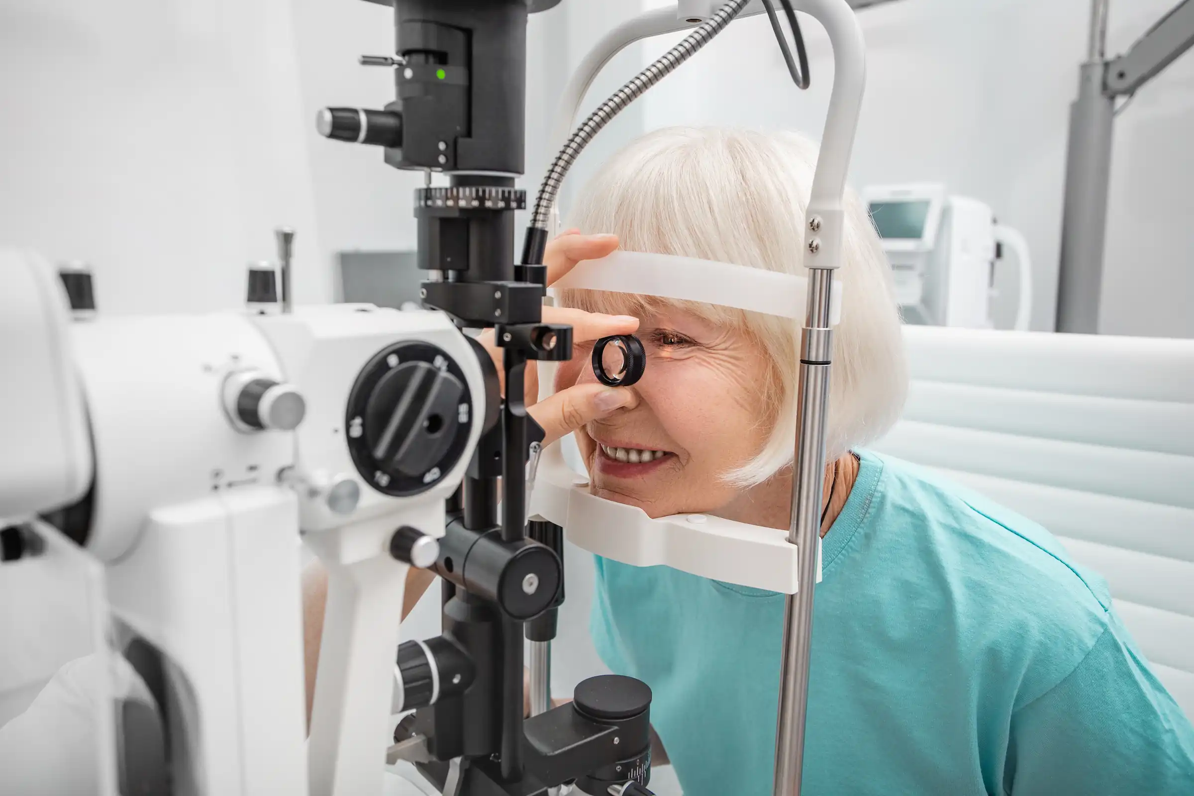

At Retina Specialty Institute, we believe knowledge is power. We never quit learning, and we work hard to educate our patients. We believe that the more you know, the more you are empowered to take charge of your health.
Learn below how to keep your eyes, particularly the retinas, healthy and protected against common diseases of the eye.
The Eye and Retina
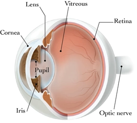
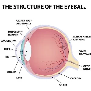
The human eye is a truly remarkable, complex machine. No other organ requires the exact operating precision of more than 30 individual parts, six muscles, and a network of instantaneous chemical connections. It’s a system of detecting the slightest movements, the furthest distances, the deepest depths, and over two million shades of color.
The retina is a thin light sensitive tissue that lines the back wall of the eye. It includes the macula, a structure that appears as a small dot inside the retina. Perhaps the most delicate of all parts of the eye, it is susceptible to damage, disease, and degeneration. Early recognition and treatment of these conditions by a highly skilled retinal specialist can maintain the health of the retina and preserve vision in most patients.
Facts about Retinal Diseases
A healthy retina is essential for good vision. Retinal diseases can affect your central vision and, if not treated, may cause partial or total loss of sight.
The three main types of retinal diseases are:
- retinal detachment
- diabetic retinopathy
- age-related macular degeneration
Retinal Detachment
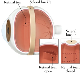
The figure above is a diagram of your eye when you experience retinal detachment. A retinal detachment occurs when the retina separates from its attachments to the underlying eye tissue. If the vitreous gel (a clear gel located in front of the retina) pulls loose from its attachment to the retina, it may cause a tear in the retina allowing the vitreous gel to pass through the tear and accumulate behind the retina. Fluid building up behind the retina causes a separation or detachment from the back of the eye.
Diabetic Retinopathy
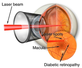
The figure above demonstrates one type of treatment for diabetic retinopathy. Diabetic retinopathy is the result of changes in the blood vessels of the retina. Blood vessels may swell, leak fluid, or grow abnormally on the surface of the retina. Because symptoms are not always noticeable in the early stages of diabetic retinopathy, annual retina screenings are recommended to help detect early warning signs and prevent long-term damage.
Age-Related Macular Degeneration
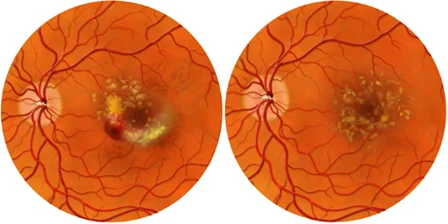
The figure above compares two different types Age-Related Macular Degeneration (ARMD), the gradual decline of vision with regards to seeing the fine details of objects. This often affects our ability to read and drive. ARMD may be a slow process that is largely unnoticeable due to small changes over time or may progress aggressively, resulting in total loss of vision.
Glossary
- Age-Related Macular Degeneration (AMD): A condition that damages the central retina, known as the macula.
- Diabetic Retinopathy: A progressive disease, affecting those with advanced diabetes, in which high sugar levels cause retinal blood vessel damage.
- Floaters: Particles or specks that appear in your line of vision.
- Macula: A small area in the center of the retina that is needed for fine visual acuity.
- Macular Edema: The swelling or thickening of the macula, caused by fluid leaking from damaged retinal blood vessels.
- Macular Hole: An abnormal opening that forms at the center of the macula over a period of several weeks to months, resulting in loss of central vision.
- Macular Ischemia: Blurred vision caused by the inadequate blood supply to the macula.
- Macular Pucker: A condition that occurs when an Epiretinal Membrane or Posterior Vitreous adheres to the macula and contracts, causing distortion or puckering of the macula.
- Retina: A nerve layer at the back of your eye that senses light and sends images to the brain.
- Retinal Detachment: Condition of the retina pulling away from its normal position.
- Vitreous Gel: A clear gel that fills the inside of the eye.
- Uvea: A layer of tissue composed largely of the blood vessels that supports and nourishes many important parts of the eye.
- Uveitis: Inflammation within the eye.
Contact Retina Specialty Institute
Inherited retinal diseases require the treatment of a highly experienced specialist. At Retina Specialty Institute, our practice is dedicated to the treatment of all forms of retinal diseases and disorders. Our experts have deep experience in the diagnosis and management of inherited retinal diseases.
The doctors at Retina Specialty Institute have either authored or reviewed and approved this content.
Page Updated: