
The eye doctors at Retina Specialty Institute offer high-quality eye care for patients in the Gulf Coast of Alabama, Mississippi, and Florida. Our retina specialists are experienced in treating a wide range of eye conditions, including retinal vein occlusion.
What Is a Retinal Vein Occlusion?
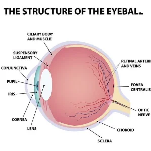
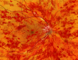
Retinal Vein Occlusion (RVO) is a serious eye condition that occurs when the veins carrying blood away from the retina become blocked. This blockage can lead to a buildup of pressure in the retina, causing vision problems and potentially leading to severe vision loss. The retina is crucial for vision as it converts light into neural signals for the brain to interpret. When these veins are blocked, it can affect this process, leading to various visual disturbances.
What Are the Types of Retinal Vein Occlusion?
Retinal Vein Occlusion is categorized into
 Trusted Source
Retina
Cleveland Clinic
Go to Source
two main types
:
Trusted Source
Retina
Cleveland Clinic
Go to Source
two main types
:
Central Retinal Vein Occlusion (CRVO):
This type involves the blockage of the main retinal vein. It is more severe and can lead to significant vision loss.
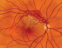
Branch Retinal Vein Occlusion (BRVO):
This occurs when one of the smaller branches of the retinal vein gets blocked. The symptoms are often less severe compared to CRVO but can still affect vision.

Retinal Vein Occlusion Symptoms
The symptoms of Retinal Vein Occlusion can vary depending on the type and severity of the blockage. Common symptoms include:
- Sudden vision loss or blurring in one eye
- Distorted vision (seeing straight lines as wavy)
- Floaters (small dark spots or lines in your vision)
- Pain or pressure in the eye (rare)
If you experience any of these symptoms, it is crucial to seek immediate medical attention
Who Is At Risk for a Retinal Vein Occlusion?
Several factors can increase the risk of developing
Trusted Source
Retina
J Clin Med.
Go to Source
developing Retinal Vein Occlusion
:
- Age: People over 50 are at higher risk.
- Hypertension: High blood pressure can damage blood vessels, increasing the risk.
- Diabetes: Diabetic patients are more prone to blood vessel damage.
- High Cholesterol: Elevated cholesterol levels can lead to atherosclerosis, impacting blood flow.
- Smoking: Nicotine use can damage blood vessels and increase blood pressure.
- Glaucoma: Elevated eye pressure can affect retinal blood flow.
Regular eye exams can help prevent vision loss due to retinal vein occlusion by enabling early detection and timely treatment of the condition, reducing the risk of severe complications. Early intervention can help manage underlying risk factors and monitor retinal health closely.
Treatments for Retinal Vein Occlusion
There is no cure for Retinal Vein Occlusion, but there are treatments available to help manage the condition and prevent further vision loss:
- Intravitreal Injections: Medications injected into the eye can reduce swelling and improve vision.
- Laser Therapy: Laser treatments can seal leaking blood vessels and reduce swelling.
- Surgical Procedures: In some cases, surgical interventions may be necessary to alleviate the blockage or address complications.
Frequently Asked Questions about Retinal Vein Occlusion
Can Retinal Vein Occlusion lead to complete blindness?
While RVO can cause significant vision loss, complete blindness is rare with prompt treatment and management.
Is Retinal Vein Occlusion painful?
Most cases of RVO are painless. However, some individuals may experience discomfort or a feeling of pressure in the eye.
Can lifestyle changes help prevent or manage Retinal Vein Occlusion?
Yes, maintaining a healthy lifestyle by managing blood pressure, diabetes, and cholesterol, quitting smoking, and regular eye check-ups can significantly reduce the risk.
How is Retinal Vein Occlusion diagnosed?
An eye doctor can diagnose RVO through a comprehensive eye exam, including imaging tests like Optical Coherence Tomography (OCT) and Fluorescein Angiography.
Is Retinal Vein Occlusion a common condition?
RVO is relatively common, especially among older adults and those with underlying health conditions. Worldwide, more than
Trusted Source
Retina
Ophthalmology
Go to Source
16 million people are affected by RVO each year
.

Schedule a Consultation
At Retina Specialty Institute, our top priority has always been and will always be offering exceptional eye care to our patients in a safe and professional environment. Contact us today for an appointment. We have a number of convenient, state-of-the-art locations.
1 Cleveland Clinic. Retinal Vein Occlusion (RVO). Available: https://my.clevelandclinic.org/health/diseases/14206-retinal-vein-occlusion-rvo Accessed July 5, 2024.
2Terao R, Fujino R, Ahmed T. Risk Factors and Treatment Strategy for Retinal Vascular Occlusive Diseases. J Clin Med. 2022 Oct 27;11(21):6340. doi: 10.3390/jcm11216340. PMID: 36362567; PMCID: PMC9656338.
3Rogers S, McIntosh RL, Cheung N, Lim L, Wang JJ, Mitchell P, et al. The prevalence of retinal vein occlusion: pooled data from population studies from the United States, Europe, Asia, and Australia. Ophthalmology. 2010;117 (2:313–319.
The doctors at Retina Specialty Institute have either authored or reviewed and approved this content.
Page Updated: